
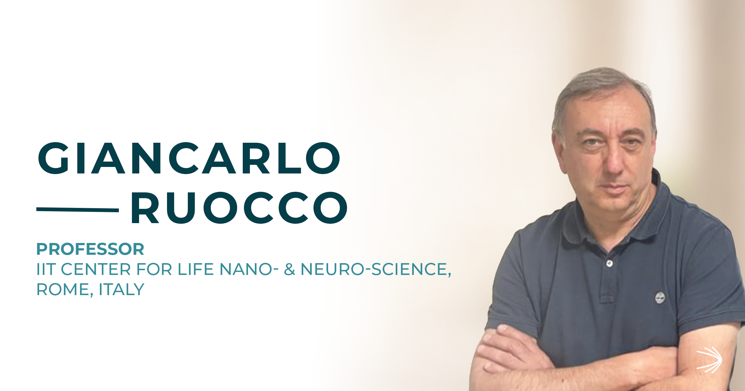
Interview with Prof. Giancarlo Ruocco
IIT Center for Life Nano- & Neuro-Science (CLN2S@Sapienza), Rome, Italy
Introduction
Every living cell has a physical character as well as a chemical one: some of its parts are stiff, others soft and fluid, a bit like the difference between a firm gel and water. These mechanical properties are not fixed; they change as a cell grows, moves, divides, and as it falls ill. Yet for a long-time researchers could see the structures inside a cell far more easily than they could measure how stiff or soft those structures were, at least not without touching or damaging the cell.
This is where Brillouin Microscopy comes in: a way of using light to “feel” the mechanical properties of living tissue, gently and without any contact. In the conversation that follows, Prof. Giancarlo Ruocco and his team at IIT in Rome describe how, together with CrestOptics, they are adding this “mechanical dimension” to everyday optical microscopy, and why it could matter for understanding diseases such as ALS. The sections below go into technical detail, but the underlying idea is simple: looking at a cell and, at the same time, sensing how soft or rigid each of its parts is.
Discovering the IIT Center for Life Nano- & Neuro-Science (CLNS)
The IIT Center for Life Nano- & Neuro-Science (CLN2S@Sapienza), established by the Istituto Italiano di Tecnologia in Rome, is an interdisciplinary research hub combining biology, physics, engineering, and nanotechnology and offering an ideal environment for innovative and translational research. Its CLN2S Imaging Facility gives researchers access to a range of advanced microscopy platforms, including spinning disk confocal, multiphoton, and super-resolution systems. These platforms support applications from single-cell dynamics to tissue imaging.
The Evolution of the IIT & CrestOptics Joint-Lab
The long-standing Joint-Laboratory between IIT CLNS and CrestOptics represents a successful model of collaboration between academia and industry. Over the years, this partnership has developed into a dynamic ecosystem where scientific curiosity, technological expertise, and industrial excellence converge. By combining the advanced biological and imaging research carried out at CLNS with CrestOptics’ engineering know-how, the Joint-Lab has progressively fostered the development and refinement of innovative microscopy solutions. This collaboration has evolved step by step, with scientific insights directly informing technological design and engineering innovation enabling new experimental possibilities. Through this continuous exchange, IIT and CrestOptics have jointly pushed the boundaries of advanced imaging technologies, strengthening the bridge between fundamental research and practical instrumentation.
“The IIT-CrestOptics Joint-Lab is a dynamic ecosystem where scientific curiosity and engineering excellence converge to push the boundaries of advanced imaging.”
Scientific Recognition within the Brillouin Technology Community
The continuous technological advancements emerging from the IIT–CrestOptics collaboration have also stimulated new scientific perspectives in the field of mechanobiology: the study of how physical forces and stiffness shape the behaviour of living cells. In particular, the development and dissemination of CrestOptics’ Brillouin Light Scattering Microscopy System, a technique that uses light to measure how stiff or soft a sample is, without ever touching it, is attracting increasing international recognition within the advanced imaging community.
This growing global traction reflects the strong potential of Brillouin technology to become a transformative tool for life-science research. By enabling the non-invasive mapping of mechanical and elastic properties of living cells and tissues, Brillouin microscopy introduces an entirely new “mechanical dimension” to conventional optical imaging. The integration of structural, molecular, and biomechanical information opens unprecedented opportunities to investigate how physical properties regulate cellular behavior, tissue organization, and disease progression. As a result, Brillouin technology is poised to significantly expand our understanding of cell biology and mechanobiology, paving the way for innovative applications in both basic and translational research.
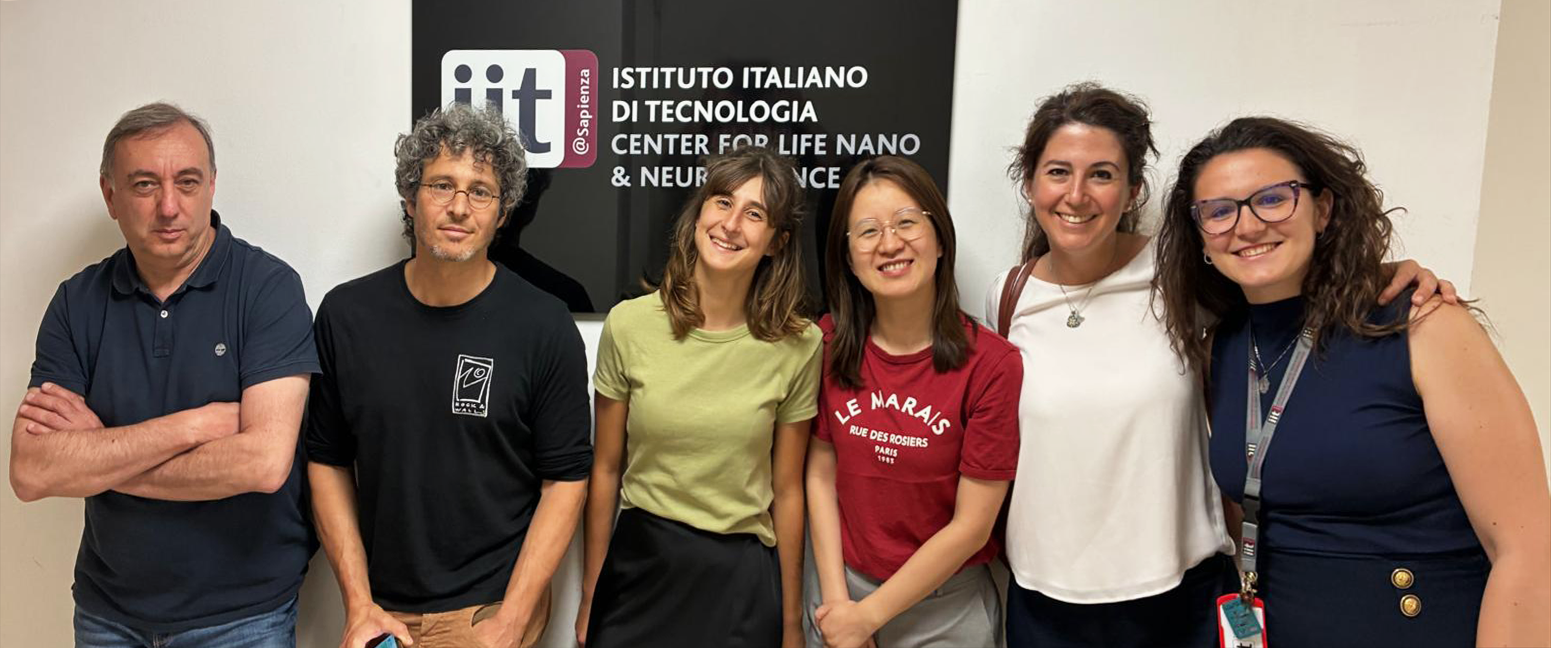
The Brillouin Research Team at IIT, led by Prof. Giancarlo Ruocco
Technological Innovation and the Brillouin Microscope Configuration
A major outcome of the IIT–CrestOptics Joint-Laboratory has been the development of an advanced Brillouin Microscopy platform specifically designed to overcome the traditional limitations of this technology in terms of acquisition speed, spatial resolution, and compatibility with live-cell imaging. The microscope’s innovative physical configuration, including its integration with the high-performance X-Light V3 Spinning Disk Microscopy system and a heterodyne detection scheme currently under development (a highly sensitive method for reading very faint optical signals), enables correlative multimodal imaging that combines mechanical information with conventional fluorescence-based cellular observations.
In other words, overlaying the “stiffness” map onto the familiar fluorescence image of the very same cell. This unique approach significantly enhances the applicability of Brillouin Microscopy to complex biological systems and dynamic mechanobiological processes. Importantly, the technology developed within the Joint-Lab has now reached a high level of maturity and is ready for translation toward the broader scientific and biomedical market. Its development has been sustained through a synergistic combination of industrial investments from CrestOptics and support from the European Pathfinder Open project ivBM. Securing such highly competitive European funding further validates scientific excellence, technological originality, and transformative potential of this imaging modality for future applications in both fundamental research and biomedicine.

Multimodal Imaging Platform at IIT, Rome: Brillouin Light Scattering & X-Light V3 Spinning Disk.
Technological Innovation and the Brillouin Microscope Configuration
A major outcome of the IIT–CrestOptics Joint-Laboratory has been the development of an advanced Brillouin Microscopy platform specifically designed to overcome the traditional limitations of this technology in terms of acquisition speed, spatial resolution, and compatibility with live-cell imaging. The microscope’s innovative physical configuration, including its integration with the high-performance X-Light V3 Spinning Disk Microscopy system and a heterodyne detection scheme currently under development (a highly sensitive method for reading very faint optical signals), enables correlative multimodal imaging that combines mechanical information with conventional fluorescence-based cellular observations.
In other words, overlaying the “stiffness” map onto the familiar fluorescence image of the very same cell. This unique approach significantly enhances the applicability of Brillouin Microscopy to complex biological systems and dynamic mechanobiological processes.
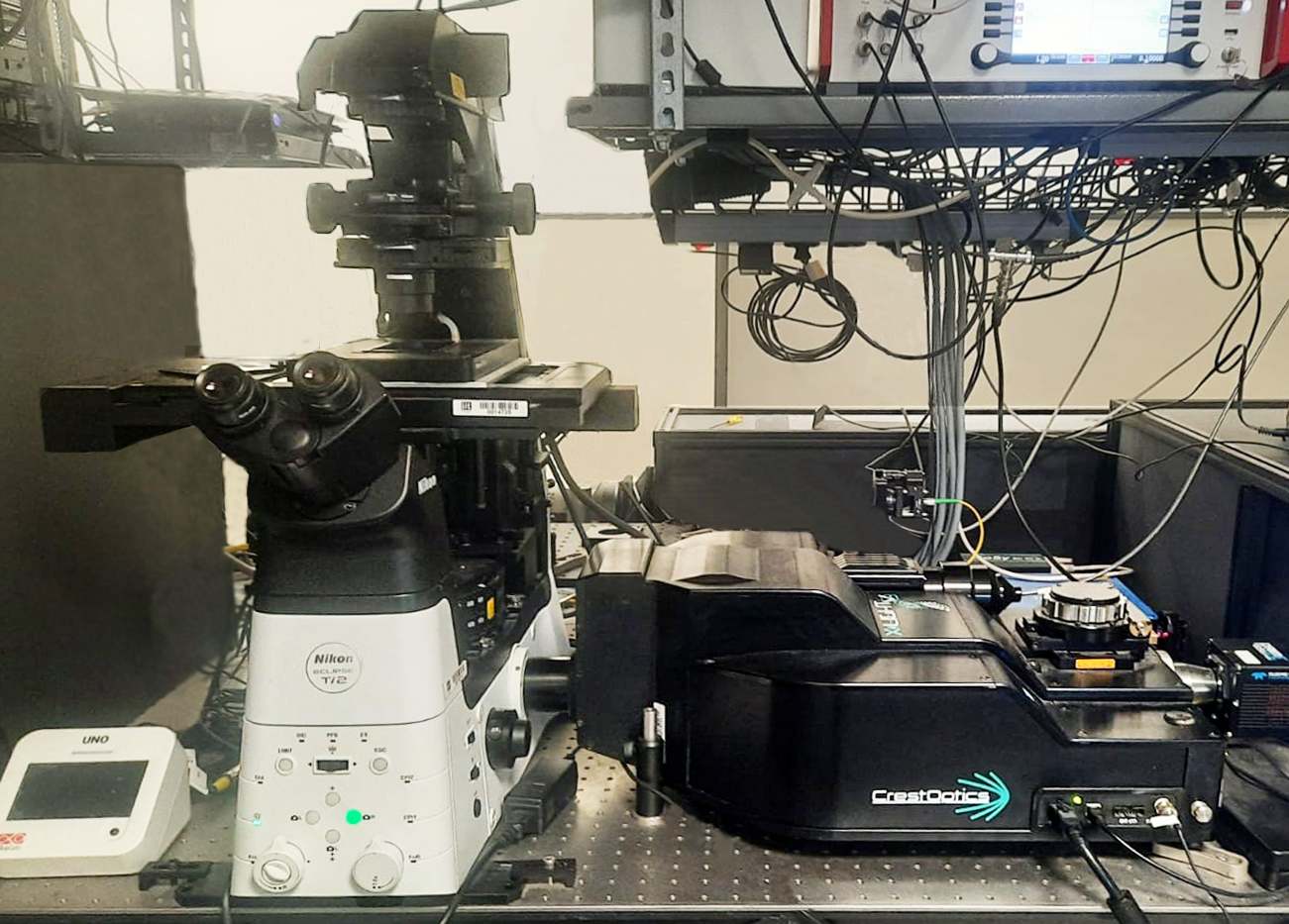
Multimodal Imaging Platform at IIT, Rome: B-Light Brillia™ Brillouin Light Scattering system & X-Light V3 Spinning Disk system.
Importantly, the technology developed within the Joint-Lab has now reached a high level of maturity and is ready for translation toward the broader scientific and biomedical market. Its development has been sustained through a synergistic combination of industrial investments from CrestOptics and support from the European Pathfinder Open project ivBM. Securing such highly competitive European funding further validates scientific excellence, technological originality, and transformative potential of this imaging modality for future applications in both fundamental research and biomedicine.
Case Study
Correlative Spinning-Disk and Brillouin Microscopy for Subcellular Mechanobiology
A compelling real-world proof of concept for the integration of CrestOptics Spinning Disk and Brillouin Microscopy technologies is provided by the recent Nature Communications publication titled “Stabilized real-time Brillouin Microscopy reveals fractal organization of protein condensates in living cells” (Testi et al., 2026). At IIT, they developed an advanced Brillouin Microscopy platform capable of performing long-term, highly stable biomechanical measurements in living cells, overcoming one of the major limitations traditionally associated with Brillouin-based imaging: temporal instability and the need for frequent manual recalibration.
They applied Brillouin Microscopy to living SK-N-BE cells, a neuroblastoma cell line commonly used as a cellular model for neurodegenerative diseases, probing protein condensates, tiny, droplet-like clusters of proteins that form inside the cell.
Specifically, the team focused on key proteins closely linked to severe neurological diseases like Amyotrophic Lateral Sclerosis (ALS) and frontotemporal dementia. They investigated the SOD1 protein, including a particularly aggressive genetic variant, alongside the TDP-43 protein and its specific fragments, which are frequently found in patient brain tissue and are believed to drive abnormal protein aggregation. To actually see these proteins in action inside living cells, the researchers tagged them with a fluorescent protein (GFP). By triggering the production of these proteins they could visually track exactly how and where they clump together within the cell, allowing precise identification of condensate formation in living cell.
To reproduce disease-relevant conditions, cells were exposed to sodium arsenite, inducing stress granule formation and promoting the assembly of pathological protein condensates. All Brillouin measurements were performed in living cells under controlled environmental conditions, using a top-stage incubation system integrated into their platform.
Within this framework, the combination of CrestOptics Spinning Disk fluorescence imaging and Brillouin Microscopy allowed direct spatial co-registration of structural and mechanical information at the subcellular level. This multimodal approach allowed the team to identify significant differences in the mechanical properties of physiological versus pathological condensates. Brillouin maps revealed significantly elevated frequency shifts in condensates formed by SOD1A4V and TDP-43 C-terminal fragments under stress conditions, consistent with increased local stiffness and reduced molecular mobility associated with pathological aggregation and cytotoxicity. In contrast, G3BP1-positive stress granules exhibited lower Brillouin shifts, reflecting their more liquid-like, dynamic nature, while GFP-only controls confirmed the absence of artefacts linked to fluorescent tagging. The resulting mechanical signatures provide experimental evidence of altered internal organization in pathological protein assemblies in living cells, with representative Brillouin maps and shift distributions shown in Figure 1.
Beyond this specific application, the work highlights how the integration of CrestOptics imaging solutions with Brillouin Microscopy can provide a powerful framework for mapping intracellular mechanics, studying biomolecular phase transitions, and generating quantitative biomechanical data that is difficult obtain with conventional imaging techniques alone.
“Integrating Spinning Disk imaging with Brillouin Microscopy provides a powerful framework for mapping intracellular mechanics and generating quantitative biomechanical data that is otherwise difficult to obtain alone.”
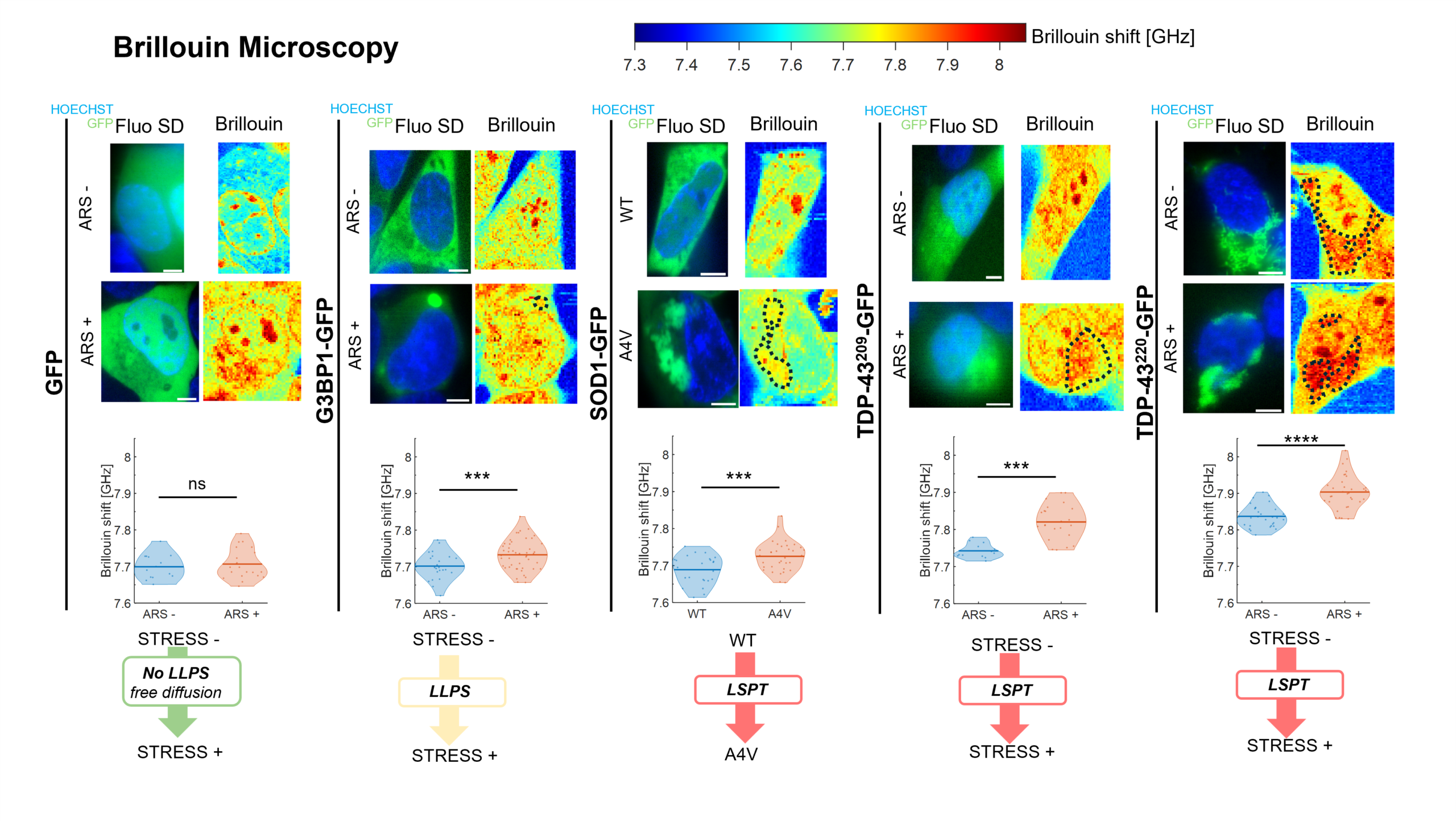
Figure 1. Brillouin Microscopy acquisitions on living SK-N-BE cells expressing different physiological and pathological biomolecular condensates. Examples of Brillouin shift maps of cells overexpressing the indicated proteins under different conditions: untreated (ARS-), treated with sodium arsenite (ARS+), or overexpressing the malignant A4V mutation (A4V). Alongside the Brillouin maps, corresponding fluorescence images display nuclei (live-stained with Hoechst) and GFP-labeled proteins. Dashed black contours indicate the location of granules within the Brillouin maps. Scale bars = 5 µm. In lower panel are reported the violin plots summarizing the distribution of Brillouin shifts for condensates in each condition, where solid lines indicate the mean of the distributions. Data were obtained from n ⩾ 3 independent biological replicates (with at least 5 cells acquired per replicate). Brillouin shifts were calculated for each cell by averaging over a binary mask derived from the GFP fluorescence signal.
Recent Related Scientific Publications from the IIT Lab Featuring CrestOptics Brillouin Microscopy Technology
Testi, C; Pontecorvo, E; Bartoli, C; Marzaro, C; Gala, F; Zhang, L; Zanini, G; D’Abbondanza, N; Garone, MG; de Turris, V; Giuliani, A; Di Timoteo, G; Bozzoni, I; Rosa, A; Ruocco, G; Nature Communications, 2026; 17(1):2387. DOI: https://doi.org/10.1038/s41467-026-68984-2.
(ii) Real-Time Brillouin Microscopy for Linewidth Imaging of protein condensates in living cells.
Bartoli, C; D’Abbondanza, N; Gala, F; Marzaro, C; Pontecorvo, E; Ruocco, G; Zanini, G; Zhang, L; Garone, M; Turris, V; Giuliani, A; Timoteo, G; Bozzoni, I; Rosa, A; Testi, C; IEEE Photonics Journal, 2026; 18 (2). DOI: 10.1109/JPHOT.2026.3655716.
(iii) Consensus statement on Brillouin light scattering microscopy of biological materials.
Bouvet, P; Bevilacqua, C; Ambekar, Y; Antonacci, G; Au, J; Caponi, S; Chagnon-Lessard, S; Czarske, J; Dehoux, T; Fioretto, D; Fu, Y; Guck, J; Hamann, T; Heinemann, D; Jähnke, T; Jean-Ruel, H; Kabakova, I; Koski, K; Koukourakis, N; Krause, D; La Cavera III, S; Landes, T; Li, J; Mahmodi, H; Margueritat, J; Mattarelli, M; Monaghan, M; Overby, DR; Perez-Cota, F; Pontecorvo, E; Prevedel, R; Ruocco, G; Sandercock, J; Scarcelli, G; Scarponi, F; Testi, C; Török, P; Vovard, L; Weninger, WJ; Yakovlev, V; Yun, SH; Zhang, J; Palombo, F; Bilenca, A; Elsayad, K.; Nature Photonics, 2025; 19, 681-691. DOI: https://doi.org/10.1038/s41566-025-01681-6.
Explore major discoveries made with our systems on our Publications page!