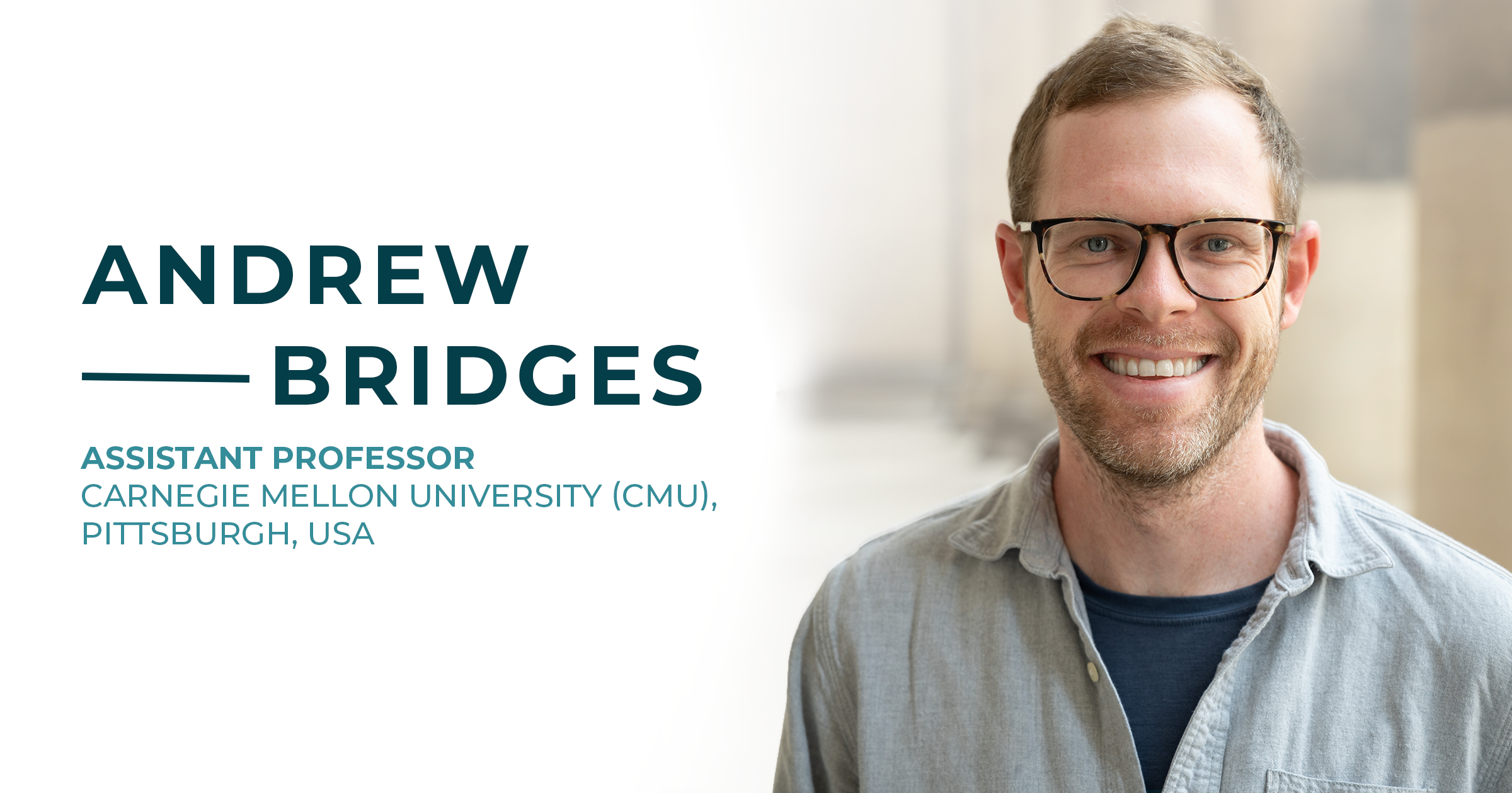
Interview with Prof. Andrew Bridges (Assistant Professor)
Carnegie Mellon University (CMU), Bridges Lab, Pittsburgh, USA
Introduction of the Carnegie Mellon University
Located in Pittsburgh, Pennsylvania, Carnegie Mellon University (CMU) is a world-renowned private research institution celebrated for its innovation in science, engineering, and technology. Consistently ranked among the top universities globally, CMU fosters a collaborative environment where interdisciplinary research thrives.
The Department of Biological Sciences at CMU is at the forefront of modern life sciences, combining computational rigor with advanced experimental techniques to solve complex biological challenges.
Advanced Research at the Bridges Lab
The Bridges Lab at Carnegie Mellon University, led by Prof. Andrew Bridges, specializes in deciphering the spatial and temporal dynamics that govern bacterial social life. The lab’s research focuses on how individual microbes transition into complex, coordinated communities known as biofilms, which provide bacteria with enhanced protection and environmental resilience.
To capture these intricate processes, the team employs cutting-edge confocal imaging. The integration of high-speed confocal systems is essential to their research, as it allows them to visualize the rapid internal dynamics of developing biofilms with high contrast and minimal phototoxicity. This setup enables the real-time observation of bacterial movement, division, gene expression, and interaction within large 3D volumes at unprecedented resolution.
A central pillar of their work is the study of how cell-cell signaling controls biofilm development, and how resulting biomechanical changes drive biofilm architecture. By decoding these fundamental “rules of engagement,” the Bridges Lab aims to uncover the molecular mechanisms of bacterial interaction and identify new vulnerabilities in these communities. Ultimately, this research provides essential knowledge to disrupt biofilm formation and address the global challenge of antibiotic-resistant infections in both clinical and environmental settings.
“The X-Light V3 is essential to our research; its high-speed volumetric imaging and sensitivity allow us to resolve the fine details of single-cell signaling within dense biofilms, capturing rapid signals and bacterial interactions that were previously difficult to visualize.”
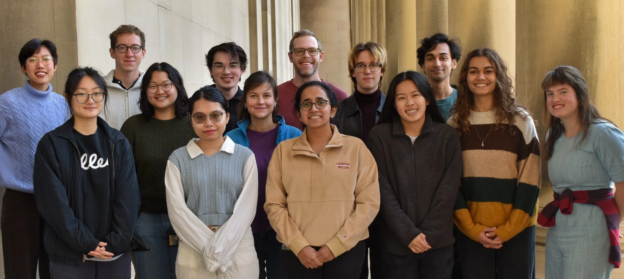
The Bridges Lab Research Team
Microscope Configuration: Setup Integrating the X-Light V3
The imaging platform at the Bridges Lab is centered on a CrestOptics X-Light V3 Spinning Disk Confocal System integrated into a motorized Nikon Ti-2E inverted microscope stand.
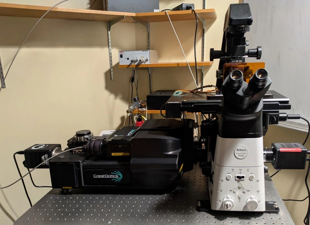
Microscope set-up at the Bridges Lab, Carnegie Mellon University, Pittsburgh
This configuration is optimized for high-performance spinning disk confocal timelapse imaging, utilizing an LDI-7 Laser Diode Illuminator (89-North) as the multi-line excitation source. To achieve maximum sensitivity and spatial resolution, the system is outfitted with a 100x silicone immersion objective (Nikon Plan Apochromat, NA 1.35) and a back-thinned Hamamatsu Orca Fusion BT sCMOS camera. This combination allows for high-speed, high-fidelity data acquisition with minimal phototoxicity, essential for longitudinal live-cell assays. All hardware components, data acquisition, and subsequent image processing are unified through Nikon NIS-Elements software, providing a streamlined and intuitive research workflow.
“The Bridges Lab Imaging Platform combines high-fidelity data acquisition with minimal phototoxicity, creating a streamlined research workflow essential for our long-term live-cell assays.”
Case Studies
1) Jojo Prentice: Cell Lysis Sensing and Biofilm Formation
Studying bacterial systems presents a significant imaging challenge due to the minute scale of the subjects; individual bacteria typically measure only 0.5 to 2.0 µm in length. Achieving the resolution required to observe signaling and behavior at the single-cell level, particularly in densely packed biofilm communities, demands an imaging pipeline capable of resolving fine details within these crowded environments. The CrestOptics X-Light V3 provides the necessary precision, allowing for the study of how lysis-sensing drives biofilm formation with remarkable clarity.
By leveraging the system’s high-speed volumetric imaging and extreme sensitivity, researchers can capture the rapid release of signaling molecules (“danger signals“) and the subsequent behavioral shifts in surrounding bacterial populations (Figure 1). The X-Light V3’s ability to maintain high temporal and spatial resolution is crucial for tracking these fast-paced, live-cell interactions in 3D, while simultaneously providing a large field of view to monitor the collective organization of the developing biofilm.
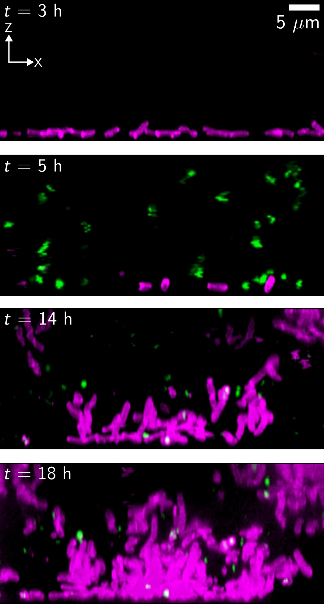
Figure 1. Left: X-Light V3 images for the indicated timepoints of Vibrio cholerae grown with a lytic bacteriophage. Magenta represents live cells expressing a constitutive reporter and green represents dead cells stained with SYTOX. After cell killing occurs, surviving cells grow back as three dimensional biofilm communities. Axes and scale bar are as indicated in the top panel. Right: X-Light V3 timelapse showing cross-sectional view of the Vibrio cholerae biofilm response to phage-mediated lysis. Live-cell signal from the constitutively expressed dL5-MG-2p complex is false-colored in magenta and SYTOX dead‐cell signal is green.
"By maintaining high spatial and temporal resolution, the system enables us to track fast-paced, live-cell interactions in 3D, revealing with remarkable clarity how processes like lysis-sensing drive biofilm formation."
2) Sandhya Kasivisweswaran: Differentiation and Post-Dispersal Cell Fate
Bacterial biofilms are highly dynamic structures where cells must constantly transition between sedentary and motile states. Understanding the mechanisms behind biofilm dispersal, and specifically the fate of the cells that remain behind, requires the ability to track individual cells over long durations across large, three-dimensional volumes. The CrestOptics X-Light V3 is uniquely suited for this task, providing the high-speed volumetric imaging necessary to capture these transition events in real-time.
By utilizing the system’s expansive field of view and rapid acquisition rates, researchers can monitor the heterogeneity within the biofilm during dispersal events (Figure 2). Ongoing work focuses on characterizing the cell fate differentiation of the population that remains in the biofilm after dispersal has occurred. The X-Light V3’s minimal phototoxicity is essential for these longitudinal studies, ensuring that cellular differentiation results from natural biological signaling rather than light-induced stress. This high-resolution temporal data allows for the precise mapping of genetic and behavioral shifts at the single-cell level within the complex architecture of the remaining biofilm; this capability also allows the Bridges Lab to interrogate the biofilm matrix components that hold cells together in these dense communities (Figure 3).
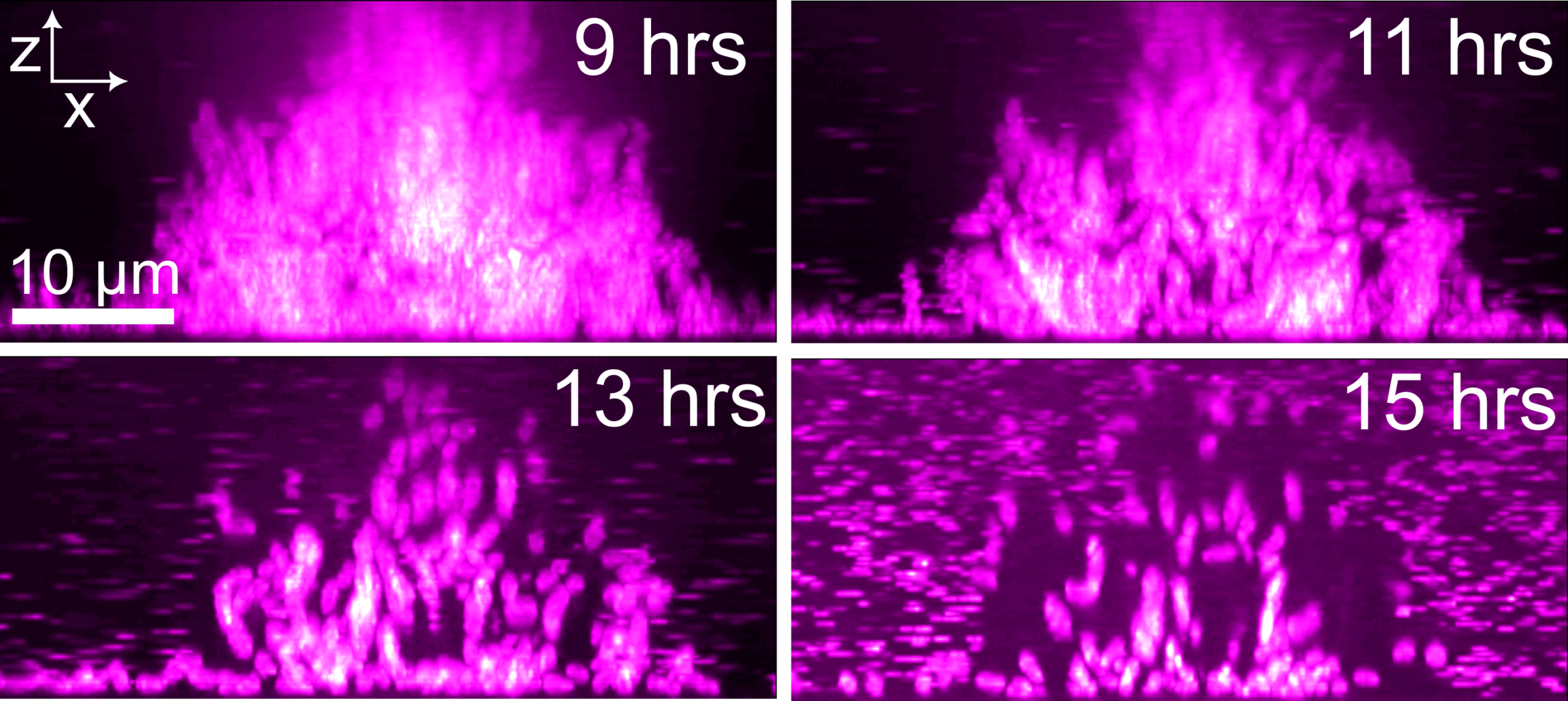
Figure 2. Left: side-on (x-z) view of high-resolution confocal micrographs of Vibrio cholerae biofilm cells expressing a constitutive reporter over the course of biofilm dispersal. The entire biofilm was optically sectioned for 16 h with 10-min intervals. Right: X-Light V3 timelapse showing WT Vibrio cholerae biofilm dispersal. Left panel: representative x-y slice. Right panel: x-z projection. Scale bar and time stamp as indicated.
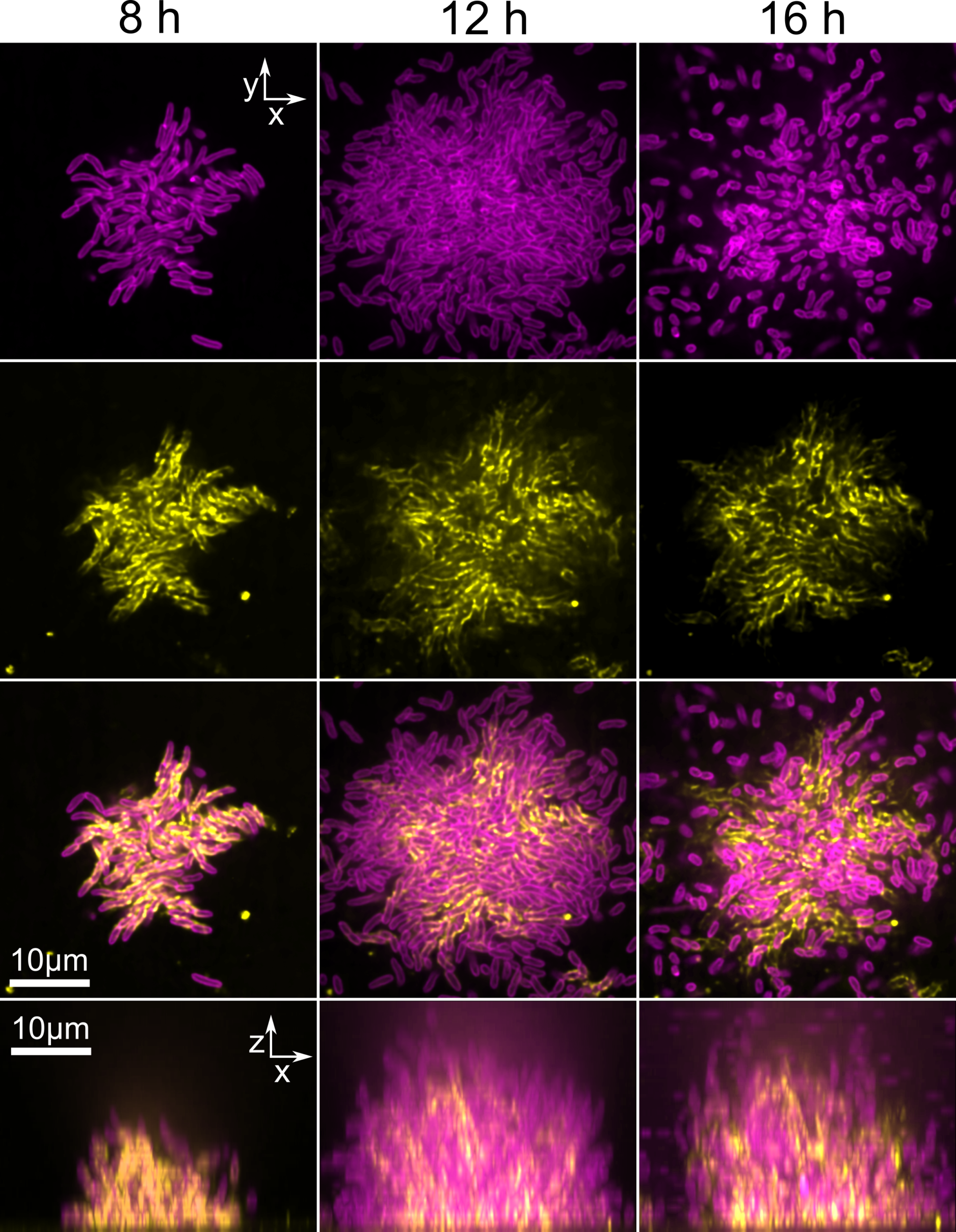
Figure 3. Left: Two color imaging of biofilm cells (magenta) and biofilm matrix components (yellow) throughout biofilm formation and dispersal. Right: X-Light V3 timelapse showing x-y projection illustrating dynamic region formation of Vibrio cholerae cells in biofilm dispersal. Arrow points to representative dynamic region. Scale bar and time stamp as indicated.
“The X-Light V3 allows us to interrogate the complex architecture of dense biofilm communities, providing the resolution necessary to visualize the matrix components that hold these bacterial societies together.”
Related Scientific Publications from the Bridges Lab Featuring Our Technology
(i) Cell-lysis sensing drives biofilm formation in Vibrio cholerae.
Jojo A. Prentice, Robert van de Weerd and Andrew A. Bridges. Nature Communications, 2024; 15(1):2018. doi: 10.1038/s41467-024-46399-1.
(ii) Biofilm dispersal patterns revealed using far-red fluorogenic probes.
Jojo A Prentice, Sandhya Kasivisweswaran, Robert van de Weerd, Andrew A Bridges. PLoS Biol, 2024; 22(11):e3002928. doi: 10.1371/journal.pbio.3002928.
(iii) A branching cell-fate decision in biofilm dispersal enables long-term surface persistence.
Sandhya Kasivisweswaran, Jojo A. Prentice, Andrew A. Bridges. BioRxiv 2026. doi: 10.64898/2026.04.24.720661.
Explore major discoveries made with our systems on our Publications page!
CrestOptics & Nikon Instruments Inc. US
We are proud of our long-term partnership with Nikon Instruments. As a key Industrial Partner, their support has been essential in bringing our technology to the global market. We would like to extend a special thanks to the Nikon team for their support in this project.