
Abstract
Brillouin microscopy is an innovative, non-invasive, and label-free optical technique that maps the 3D mechanical properties (stiffness and viscosity) of biological samples at sub-micron resolution, without physical contact or structural damage.
When integrated with spinning disk confocal fluorescence microscopy, it becomes a powerful multimodal platform: fluorescence pinpoints exact molecular structures like a GPS, while the Brillouin channel maps their biomechanical properties on the exact same focal plane.
As the first chapter in our dedicated series on Brillouin microscopy, this Application Note introduces the core physical principles of the technique, outlines its primary biomedical applications, and presents a real-world case study. Through this example, we demonstrate how a stabilized Brillouin-confocal setup reveals the fractal architecture and pathological hardening of protein condensates in live-cell models of Amyotrophic Lateral Sclerosis (ALS) and Frontotemporal Dementia (FTD).
Future parts in this series will delve deeper into technical aspects of this technique and emerging application areas of Brillouin imaging.
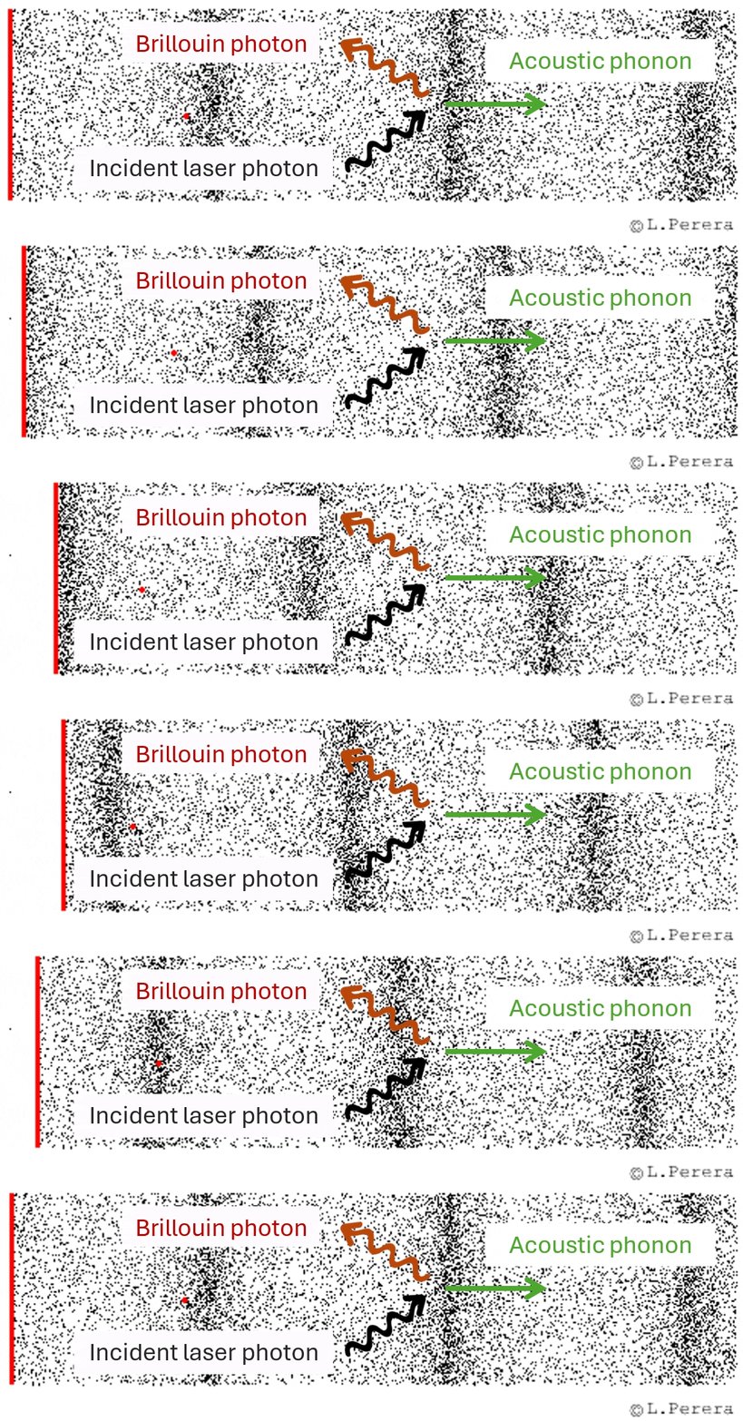
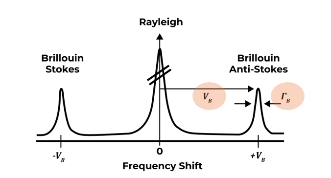
Figure 1: Working principle of Brillouin light scattering. (Top) Schematic representation of an incident photon interacting inelastically with a thermally generated acoustic phonon, giving rise to a frequency-shifted Brillouin photon. (Bottom) Representative Brillouin spectrum consisting of a central elastic Rayleigh peak and two symmetric inelastic Brillouin peaks (Stokes and Anti-Stokes). The frequency shift (νB) correlates with the material’s stiffness (longitudinal modulus), while the spectral linewidth (ΓB) reflects its local viscosity.