Interview with Prof. Paolo Netti
Department of Chemical, Materials and Industrial Production Engineering (DICMAPI), University of Naples Federico II, Italy
Center for Advanced Biomaterials for Health Care, Istituto Italiano di Tecnologia (IIT), Naples, Italy
Interdisciplinary Research Centre on Biomaterials (CRIB), University of Naples Federico II, Italy
About Prof. Paolo Netti
Paolo Netti, Professor at the Department of Chemical, Materials and Industrial Production Engineering (DICMAPI) at the University of Naples Federico II, Director of the Center for Advanced Biomaterials for Healthcare (CABHC), and Coordinator of the Bio-Logic Materials research line at the Istituto Italiano di Tecnologia (IIT) in Naples, is a chemical engineer by training. His research focuses on developing engineering principles to control and guide biological systems. His work lies at the intersection of bioengineering, materials science, mechanics, and biology, with a particular focus on understanding how the physical properties of materials can be engineered into instructive signals that shape cellular function and fate.
Over the course of his career, his research has progressively evolved from developing biomaterials as passive scaffolds toward active, instructive systems capable of directing biological organization across multiple scales. This includes the development of cell-instructive materials providing biochemical and biophysical cues, as well as engineered 3D microtissues and tissue models that reproduce key features of the cellular microenvironment.
This vision defines the research carried out by his group, which combines biomaterials engineering, mechanobiology, advanced microscopy, quantitative imaging, and cell biology. By bringing these disciplines together, the group develops engineered culture platforms and analytical approaches that connect material design with biological function, allowing biological systems to be investigated from the cellular and tissue scale down to their genome activation.
Research Institutions: UNINA & IIT Naples
The joint research infrastructure of the University of Naples Federico II and the Istituto Italiano di Tecnologia (IIT) brings together complementary expertise and state-of-the-art facilities in microfabrication, biomaterials engineering, cell culture, and advanced microscopy. This creates an integrated environment where engineered materials and in vitro biological systems can be designed, fabricated, and characterized within the same research framework.
Within this ecosystem, the Imaging Platform provides essential analytical capabilities to characterize newly developed systems and evaluate the biological responses they elicit. By employing a combination of advanced optical and electron microscopy techniques, researchers extract both qualitative and quantitative data, spanning from cellular morphology and organization to subcellular features and functional responses.
In daily practice, this environment enables a continuous, iterative workflow: researchers design engineered culturing platforms, observe cell-material interactions, quantitatively characterize cellular responses, and feed this data back to refine material design. The Imaging Platform serves as an essential analytical component of their research, helping them translate engineered microenvironments into measurable biological outcomes.
Research Focus: The Bio-Logic Materials Line
Mechanobiology has become an increasingly important component of the group’s research because cell function cannot be fully understood by considering biochemical activity alone. As Prof. Netti points out: “Cells continuously sense and respond to biophysical cues within their surrounding environment, which can shape their mechanical state and, in turn, influence cell behavior, function, and fate.”
"Cells continuously sense and respond to biophysical cues within their surrounding environment, which directly shape their mechanical state and, in turn, influence cellular function and fate."
Within this context, the group has developed extensive expertise in engineering culturing platforms that provide controlled biophysical signals to cells capable of modulating their mechanical identity. Measuring how such cues influence cellular mechanical state, ideally through non-contact and label-free approaches, therefore represents an important step in their experimental workflow.
This is precisely where Brillouin microscopy provides a particularly valuable perspective. By measuring the Brillouin shift with spatial resolution, it allows them to detect local variations in the mechanical response of a cell (Figure 1). Because the measurement is performed without physical contact, it offers the possibility of probing living biological systems without mechanically perturbing them.
Ultimately, this integration opens new opportunities to investigate how mechanical states vary across a cell, how they evolve over time, and how they are associated with changes in cellular organization and function. In combination with engineered microenvironments, Brillouin microscopy therefore provides a way to connect the physical state of a cell with the conditions that shape its behavior.
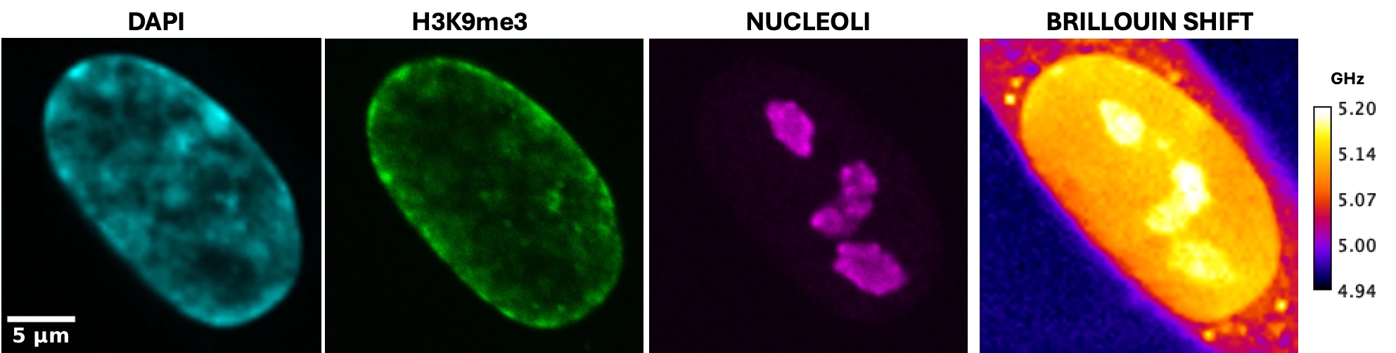
Figure 1. Correlative confocal and Brillouin shift imaging of a human adipose stem cell nucleus. Representative images showing spatial correlation between molecular markers and mechanical properties. Left to right: overall nuclear morphology (DAPI, cyan), heterochromatin marked by H3K9me3 domains (green), nucleoli marked by nucleolin (magenta), and the corresponding Brillouin frequency shift map (GHz, 4.94–5.20 GHz). Scale bar: 5 µm
The Synergy with CrestOptics
The collaboration between Prof. Netti and CrestOptics developed from a shared interest in overcoming one of the primary limitations of advanced imaging: mechanobiology increasingly requires the integration of structural, mechanical, and functional information, whereas most conventional microscopy platforms treat each as an isolated measurement. The group’s research required the ability to measure local mechanical properties and, crucially, to spatially associate these mechanical signatures with specific cellular or extracellular components. Combining Brillouin microscopy with confocal fluorescence imaging offered a powerful solution to address this need. Concurrently, CrestOptics sought to explore how the Brillouin-based imaging technology could be integrated into a broader bioengineering workflow and applied to biological questions within the group’s research line. The convergence of these scientific and technological goals provided the foundation for the collaboration.
From a scientific perspective, transitioning from a single-channel approach to a multimodal platform is essential for mechanobiology. Brillouin microscopy provides quantitative data on the local mechanical response of a region of interest, but interpreting the origin and biological significance of these mechanical signatures requires complementary context. Fluorescence microscopy adds a complementary molecular and structural dimension, allowing researchers to identify specific cellular or extracellular components and relate their organization, state or activity to the local mechanical response measured by Brillouin microscopy.
As Prof. Netti highlights: “The real advantage of combining the two modalities is therefore not simply the acquisition of more information, but the ability to establish spatial relationships between mechanical responses and biological structures within the same sample. This provides a more integrated view of how physical properties are coupled to biological organization and function across different scales.”
“The real advantage of combining these modalities isn't just getting more data, it's establishing spatial links between mechanical responses and biological structures within the same sample.”
The Integrated Brillouin Platform
The key strength of the platform lies in the tight integration of CrestOptics Brillouin Light Scattering solution with the high-performance X-Light V3 Spinning Disk Confocal system, allowing both modalities to operate within a unified optical framework.
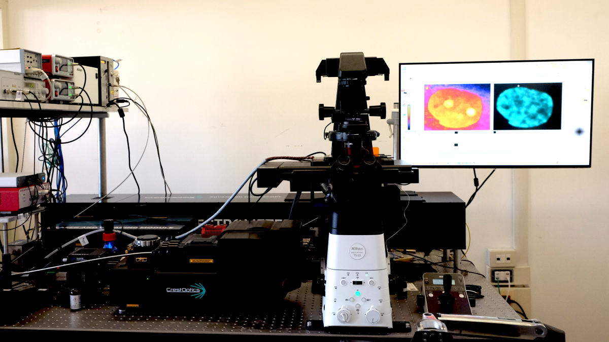
Multimodal Imaging Platform at the Interdisciplinary Research Centre on Biomaterials (CRIB), University of Naples Federico II, Naples: B-Light Brillia™ Brillouin Light Scattering system and X-Light V3 Spinning Disk system.
One of the main engineering advantages is the precise spatial alignment between the Brillouin and fluorescence channels, particularly in the x–y plane. This alignment is essential for their applications because it allows them to relate a local mechanical measurement directly to the biological structures identified by fluorescence, rather than treating the two measurements as independent datasets. Another key advantage is the ability to navigate through the sample along the z-axis using the spinning disk confocal system and perform measurements at different depths. This enables three-dimensional acquisition while maintaining strict spatial correspondence between both modalities, allowing them to construct volumetric 3D maps in which mechanical and molecular information are directly correlated.
High spatial resolution is equally critical for these applications, as closely adjacent cellular or extracellular structures often exhibit distinct mechanical properties. Spatially resolved Brillouin measurements allow researchers to detect local variations in the Brillouin signal and assign them to specific fluorescently tagged structures, avoiding signal averaging across larger regions.
Furthermore, the optical stability of the integrated platform is equally important. Brillouin measurements rely on the detection of intrinsically weak inelastic scattering signals and are therefore sensitive to optical fluctuations. Maintaining a stable and precisely aligned optical configuration is essential for obtaining accurate and reproducible measurements across different acquisitions and sample regions. The platform is supported by an integrated software environment that coordinates both imaging modalities within a streamlined acquisition workflow, enabling seamless switching, region-of-interest selection, and automated spatial registration.
In conclusion, Prof. Netti emphasizes that: “The combination of Brillouin Light Scattering with Spinning Disk Confocal Microscopy offers a promising direction for advancing our research in mechanobiology. This approach enables the simultaneous visualization of cellular structures alongside intravital measurements of their mechanical properties. While technically and conceptually demanding, such integration creates an opportunity to explore how forces and mechanical cues influence cell behavior and decision-making processes.”
Recent Related Scientific Publications from Prof. Netti’s group
(i) Substrate Curvature and Stiffness Equivalence in Governing Cell Mechanosensing
C. Frascogna, V. Panzetta, V. Mollo, R. Marotta, S. Strano, S. Fusco, P. A. Netti; Nature Communication, 2026. DOI: 10.1038/s41467-026-76866-w
(ii) Material-Induced Nuclear Deformation Controls Chromatin Architecture in Adipose Stem Cells
C.F. Natale, L. Messina, V. Panzetta, S. Saporito, C. Menna, M. Ventre, P. A. Netti; Advanced Science, 2026, DOI:10.1002/advs.202514458
F. Mauro, C. F. Natale, V. Panzetta, V. Mollo, P. A. Netti; Biomaterials, 2026, DOI: 10.1016/.biomaterials.2025.123512
Explore major discoveries made with CrestOptics systems on our Publications page!